Of all the immunochemical techniques, the Western blot is a unique method. It is the only one that uses electrophoretic separation as a starting point for analysis. Although it is time-consuming, it has found widespread use in laboratories around the world.
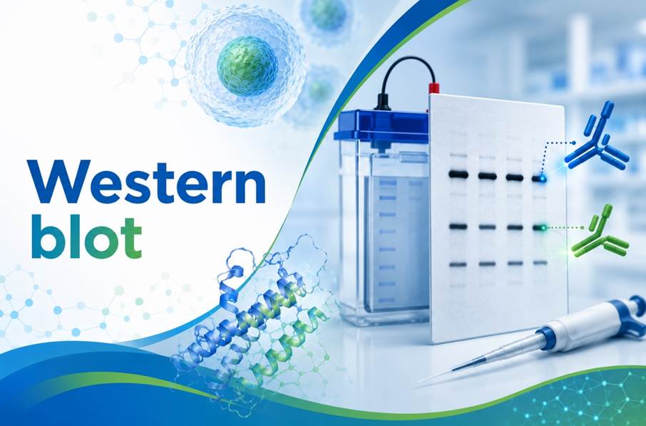
What is a Western blot?
Western blotting is a multi-step analytical technique. It is used to detect specific proteins in biological samples, such as cell lysates, tissue homogenates and body fluids.
How does a Western blot work?
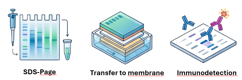
A Western blot analysis can be divided into several stages:
- Protein preparation: This stage may vary depending on the matrix being analysed. In the case of cells, lysis must be performed to extract the proteins. Sometimes it is necessary to isolate proteins from cell membranes using detergents. Some matrices may require the use of various techniques, such as dialysis, ultrafiltration, SPE, etc.
- SDS-PAGE: The isolated proteins are separated by electrophoresis. Their separation is based on molecular weight. You can find out more about SDS-PAGE in this article.
- Transfer to a membrane: proteins trapped in the pores of the gel are difficult to analyse further. The gel is fragile and impermeable to antibodies. It is therefore necessary to transfer the proteins from the gel to a membrane.
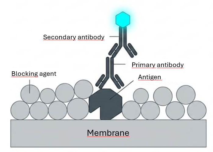
- Blocking the membrane: As with the ELISA technique, the membrane must be blocked. This prevents non-specific binding of antibodies to the membrane itself. BSA or milk proteins are most commonly used for blocking.
- Immunodetection: Labelled antibodies are used to detect proteins. These bind specifically to the target protein. Any excess antibodies must be washed away. Depending on the detection method chosen, the proteins appear as coloured or fluorescent bands.
Where does the name ‘Western Blot’ come from?
The history of the naming of blotting techniques is one of the most fascinating stories in molecular biology.
It all began in 1975, when Edwin Southern described a method for transferring DNA fragments onto a membrane. The technique was named the Southern blot, after its inventor.
Two years later, a similar method was developed for RNA; the scientists, playing on the analogy with the cardinal directions, called it the Northern blot.
In 1979, Harry Towbin and his colleagues described a method for transferring proteins, but it was W. Neal Burnette who, in 1981, coined the term “Western blot“. Burnette, who was working at the Fred Hutchinson Cancer Research Centre at the time, wanted to continue the tradition started by Southern and the “northern” variant for RNA. Although the editors of scientific journals initially rejected the name as too colloquial, the scientific community embraced it with enthusiasm
Types of protein transfer
Transfer—the process of transferring proteins from the gel to the membrane—is a critical step that determines the sensitivity and reproducibility of the entire experiment. The proteins must retain the pattern (known as the electrophoretic map) established in the gel.
Wet transfer
This is the most traditional and efficient method. The gel and membrane are sandwiched between two plates and completely immersed in a tank containing the transfer buffer.
- Advantages: Maximum transfer efficiency for high-molecular-weight proteins (>150 kDa). The system’s cooling capability allows for extended transfers at high voltages.
- Disadvantages: Requires a large amount of buffer and time (often overnight procedures).
Semi-dry Transfer
In this configuration, the ‘sandwich’ is placed directly between two flat plate electrodes. The only source of electrolyte is the buffer solution with which the filter papers surrounding the gel and the membrane are saturated.
- Advantages: Speed (15–60 minutes) and reduced reagent consumption.
- Disadvantages: Reduced efficiency with very large proteins; risk of the gel overheating if the transfer time is extended.
Dry Transfer
Modern automated systems use special gels and membranes with built-in electrolytic matrices.
- Advantages: Extremely fast (even under 10 minutes), no need to prepare buffers.
- Disadvantages: The high cost of manufacturer-specific consumables.
Types of membranes: PVDF vs Nitrocellulose
Immunodetection takes place on a special membrane. Its properties help to immobilise proteins. The immobilised proteins can then be detected by antibodies. Two types of membrane are used in Western blotting:
- Nitrocellulose: A classic choice, characterised by good protein binding and low background. However, it is fragile and is not recommended for procedures requiring repeated removal and reapplication of antibodies (stripping).
- PVDF (polyvinylidene fluoride): It exhibits significantly greater mechanical strength and a higher binding capacity for (hydrophobic) proteins. However, it requires pre-activation in methanol.
Detection methods
The Western blot method uses two types of antibodies:
- Primary antibodies – antibodies that recognise the antigen of the protein being analysed. These are most commonly rabbit or mouse antibodies. They can recognise the protein or a specific form of it, such as a phosphorylated form.
- Secondary antibodies – these are labelled antibodies that specifically recognise the Fc fragment of primary antibodies.
Secondary antibodies can be labelled with various types of markers. Depending on the laboratory’s equipment, different detection methods can be selected.
Chromogenic detection (HRP)
This method uses the enzyme HRP (horseradish peroxidase) conjugated to a secondary antibody. Upon addition of a substrate, such as TMBV (3,3′,5,5′-tetramethylbenzidine), a coloured band appears on the membrane.
Other chromogenic substrates used include:
DAB – 3,3′-diaminobenzidine – dark brown colour
4 – CN – purple-blue
AEC (3-amino-9-ethylcarbazole) – a red precipitate
Chemiluminescent detection (HRP)
The most commonly used method involves the enzyme HRP (horseradish peroxidase) conjugated to a secondary antibody. Upon addition of the substrate (luminol), the enzyme catalyses an oxidation reaction, resulting in the emission of light.
- Features: Extremely high sensitivity (femtogram level). The signal is recorded on X-ray film or in digital systems using a CCD camera. However, this is a kinetic method – the signal fades as the substrate is depleted.
Fluorescence detection
Here, secondary antibodies are labelled with fluorophores (e.g. IRDye dyes, Alexa Fluor).
- Advantages: It allows for multiplexing, i.e. the simultaneous detection of several proteins of different molecular weights on the same membrane using different wavelengths. The signal is stable and linear over a very wide range, making this method ideal for precise quantitative analysis.
Limitations of the method
The Western blot has limitations that every researcher must bear in mind:
- Antibody specificity: The result is only as reliable as the antibody used is specific. Poor-quality antibodies may produce non-specific signals (additional bands) or high background levels.
- Semi-quantitative nature: Western blots rarely provide absolute molecular counts. We usually determine relative changes (e.g. ‘expression has doubled’), which requires the use of loading controls (e.g. actin, GAPDH).
- Narrow dynamic range (in ECL): In chemiluminescence detection, signal saturation can easily occur, which prevents the reliable quantitative assessment of samples with very high protein concentrations.
- Transfer artefacts: Air bubbles between the gel and the membrane cause ‘holes’ in the band pattern.
- Protein preparation: During sample preparation for analysis, proteins may fragment or form aggregates. This can result in the appearance of multiple bands and lead to inaccurate results. It is advisable to use protease inhibitors and surfactants to prevent aggregation.
Western blot modifications
Science is evolving, and so is the Western blot. Here are some of the modifications:
- Far-Western Blot: This technique is used to study protein-protein interactions. Instead of a primary antibody, a different protein is used, which is designed to bind to the antigen on the membrane.
- Southwestern Blot: This technique is used to study protein-DNA interactions. Labelled DNA probes are applied to a membrane on which proteins have been separated.
- Native Western Blot: Omit the SDS denaturation step. This allows proteins to be analysed in their natural form, preserving their quaternary structure and protein complexes.
- Automated Western Blot (Capillary WB): Modern systems (e.g. Jess, Wes) carry out the entire process in microcapillaries, which eliminates manual errors and allows for the analysis of 24 samples in 3 hours.
- Dot blot: The protein sample is applied directly to the membrane, bypassing the electrophoresis stage. It is suitable as a screening method for the rapid detection of proteins, without quantification.
Application of Western Blot
- Diagnosis of infectious diseases: For many years, this was the ‘gold standard’ for confirming HIV and Lyme disease infections (detection of antibodies against specific viral proteins).
- Oncological research: Monitoring the levels of oncoproteins and the phosphorylation status of signalling proteins in response to novel therapies.
- Neurology: Detecting abnormal forms of proteins, such as amyloid-beta in research into Alzheimer’s disease or prions.
- Quality control of medicines: Verification of the purity of recombinant proteins and monoclonal antibodies used in medicine.
- Gene expression analysis: Western blotting is widely used to analyse gene expression at the protein level. This method is frequently employed in clinical trials to monitor the levels of selected biomarkers.
- Analysis of cellular signals: Primary antibodies can recognise specific protein sequences and even their different forms, such as phosphorylated forms. This makes it possible to study receptor activation and intracellular signal transduction (e.g. an increase in receptor phosphorylation levels in response to a stimulus)
Summary
It has been almost half a century since the Western Blot technique was developed. Despite the passing of time, it is still used in biology laboratories around the world today. Remember to use high-quality reagents, choose the appropriate detection method, and prepare your sample carefully. The quality of the results obtained depends on this.
Bibliography and sources
- Burnette, W. N. (1981). “Western blotting”: electrophoretic transfer of proteins from sodium dodecyl sulfate-polyacrylamide gels to unmodified nitrocellulose and radiographic detection with antibody and radioiodinated protein A. Analytical Biochemistry, 112(2), 195-203.
- Mahmood, T., & Yang, P. C. (2012). Western blot: technique, theory, and trouble shooting. North American Journal of Medical Sciences, 4(9), 429-434.
- Towbin, H., Staehelin, T., & Gordon, J. (1979). Electrophoretic transfer of proteins from polyacrylamide gels to nitrocellulose sheets: procedure and some applications. Proceedings of the National Academy of Sciences, 76(9), 4350-4354.
Q&A
The PVDF membrane is highly hydrophobic (it repels water). Methanol ‘wets’ the pores of the membrane, allowing the transfer buffer to penetrate the structure, which is essential for contact to occur between the proteins and the membrane.
Skimmed milk is cheaper and very effective, but it contains proteins (e.g. casein) that may interact with antibodies against phosphorylated proteins. In studies involving phosphoproteins, it is recommended to use BSA (bovine serum albumin).
This is a typical result of the gel overheating during electrophoresis. Excessive voltage generates heat, which distorts the front of the band. You should reduce the voltage or carry out the process at a lower temperature.
This is the process of removing antibodies from the membrane once detection is complete, without damaging the membrane-bound proteins themselves. This allows the same membrane to be reused for the detection of another protein (e.g. a loading control).
Proteins may undergo post-translational modifications (e.g. glycosylation) that increase their molecular weight. Furthermore, strongly basic or acidic proteins may bind SDS in an unusual way, which alters their electrophoretic mobility.