If you are reading this article, you are facing the challenge of counting cells in suspension. We have already written about different methods of cell counting. In this article, we will answer the question: How to count cells in a Bürker chamber?
What is a Bürker chamber?
A Bürker chamber is a type of haemocytometer, a special microscope slide for counting blood cells. Thanks to its special design, it enables quick and accurate counting of cells in a sample. It takes only a few minutes for experienced specialists. Counting is performed during microscopic observation.
How is a Bürker chamber constructed?

The Bürker chamber looks like a microscope slide. The slide has a special shape. It has four smooth surfaces, two central ones (top and bottom) and two side ones. The central surfaces are slightly below the side surfaces (0.1 mm). Grids are drawn on these surfaces to count cells. The cover glass rests on the side surfaces.
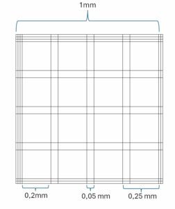
The grid drawn on the central surfaces consists of 9 large squares (surrounded by three lines). Each of these squares has an area of 1 mm² and is divided into 25 smaller squares measuring 0.2 x 0.2 mm and 25 squares measuring 0.05 x 0.05 mm. These squares are used to count cells of different sizes. For example, lymphocytes are counted in the largest squares, erythrocytes, which are much more numerous, in the medium squares, and platelets in the smallest squares.
The grid, together with the cover glass, creates a space with known dimensions. This makes it possible to accurately determine the cell density.
Preparation of cell suspension
Before counting, the cells must be properly prepared. They are usually in a small volume of medium. In this case, it is advisable to dilute the cells before placing them on a slide. Remember to move the vessel up and down each time before taking a cell sample to suspend them well. Otherwise, they will settle to the bottom of the vessel as a result of sedimentation and the measurement result will be unreliable. Approximately 10 μl of suspension is sufficient for the measurement.
Preparing the counting chamber
To prepare the chamber, place a cover glass on it so that it remains stationary. Some chambers are equipped with special clamps that hold the glass in place. However, if there are no clamps, lightly moisten the two smooth side surfaces. The cover glass can be lightly pressed down with your fingers to hold it in place. The chamber is now ready.
Trypan blue staining
Usually, when counting cells, we are only interested in the living ones. It is therefore useful to distinguish between dead and living cells and determine cell viability. This will provide us with valuable information about the condition of the culture we are growing.
Trypan blue is used to distinguish dead cells from living cells under a microscope. It penetrates dead cells and stains them blue. To stain a sample, add 10 μl of trypan blue to 10 μl of cell suspension and mix well.
WARNING! From this moment on, you must act quickly. Trypan blue is toxic and after about 5 minutes it begins to kill cells, thus distorting the result.
Counting
After adding trypan blue, we can place the sample into the Bürker chamber. It is best to prepare the chamber in advance. Using an automatic pipette (preferably 1-10 μl or 2-20 μl), we apply the stained cell suspension to the chamber. We do this by pipetting 10 μl between the cover glass and the central surface. A volume of 10 μl will be sufficient to remove air bubbles.
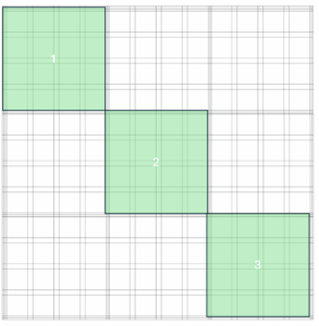
When counting, it is important to count each cell only once. For beginners, this can be a problem, especially when cells lie on the boundary lines between fields. However, there is a way around this. For each field, we select two lines (e.g. the left and top lines). Cells lying on these lines are counted as part of that square. If a cell lies on a different line, it is counted as part of the neighbouring square. This ensures that we do not count cells twice. It is also worth using clickers, so that we do not have to focus on counting and can simply click on the cells.
Calculations
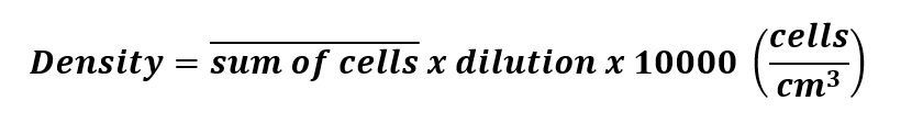
After taking the measurement, it is time for calculations. To determine the cell density, divide the measurement result by the chamber volume. This is determined by the grid dimensions and the height of the cover glass. For large cells, which we count in the largest squares, the chamber has dimensions of 1 x 1 x 0.1 mm – 0.001 cm³.

In our calculations, we should also take into account the dilution of cells that we performed when preparing the cells and the dilution of cells as a result of staining with trypan blue. The final formula after simplifying the division is as follows

NOTE! In order to reduce error, I recommend counting in at least three squares and averaging the measurement result. It is a good idea to choose squares diagonally to cover the front, middle and back of the grid.