Electrophoresis is one of the fundamental techniques used in molecular biology, biochemistry and genetics. It enables the separation, identification and purification of electrically charged molecules – mainly nucleic acids (DNA, RNA) and proteins. Electrophoresis has undergone many variations and modifications. It can also be combined with other analytical techniques such as immunometry or mass spectrometry. In this article, we will explain the principle of electrophoresis.
What is electrophoresis??
Electrophoresis is based on the ability of charged particles to move in an electric field. The name of the technique derives from this ability (electro – electricity, foresis – movement). Positively charged molecules will be attracted to the cathode, while negatively charged molecules will be attracted to the anode. This phenomenon was first observed by Alexander Reuss in 1807. In 1908, electrophoresis was used for the first time as an analytical technique. Karl Landsteiner used it to separate plasma proteins.
The migration velocity of molecules (V) depends on the electric field intensity (E), the molecular charge (q) and the friction coefficient (f), which is described by the equation:
V = Eq/f
The friction coefficient depends on the size and shape of the particle and the viscosity of the medium in which the movement occurs.
In laboratory practice, this means that:
- Particles with lower mass migrate faster.
- Particles with a higher charge are more strongly attracted to the electrode with the opposite charge.
- The medium (gel) acts as a molecular sieve, slowing down larger particles.
How is electrophoresis performed?
Electrophoretic separation is performed on a gel, which acts as a molecular sieve through which molecules migrate. Water and salts present in the gel, which is often immersed in a buffer, enable the conduction of electric current. Electrophoresis can be performed on a gel in a horizontal position (the gel is poured onto a base) or in a vertical position in a special cassette. There are also more advanced techniques such as capillary electrophoresis or microelectrophoresis in special cartridges.
Types of gels
Choosing the right medium (gel) is crucial to the success of the experiment. By selecting the gel and its density, we can control the size of the pores. The size of the pores, in turn, directly affects the resolving power.
Agarose gels
Agarose is a polysaccharide obtained from seaweed. These gels are characterised by large pores and are most commonly used in horizontal electrophoresis.
- Application: Mainly separation of nucleic acids (DNA/RNA) ranging in size from several hundred to several thousand base pairs (bp).
- Configuration: Usually horizontal electrophoresis.
Polyacrylamide gels
They are formed as a result of the polymerisation of acrylamide with a cross-linking agent (bis-acrylamide). They form a dense network with small pores.
- Application: Separation of proteins and short DNA/RNA fragments (high resolution, up to 1 bp).
- Configuration: Typically vertical electrophoresis..
How to perform electrophoresis?
A power source is required to perform electrophoresis. Special electrophoresis power supplies are used for this purpose, which allow the voltage and current to be regulated. The gel is placed in a special apparatus, whose electrodes are connected to the power supply. Both the electrodes and the gel are immersed in a buffer that allows the current to flow. During separation, the temperature rises as a result of electrical resistance and friction. For this reason, the entire system is usually equipped with a cooling system.
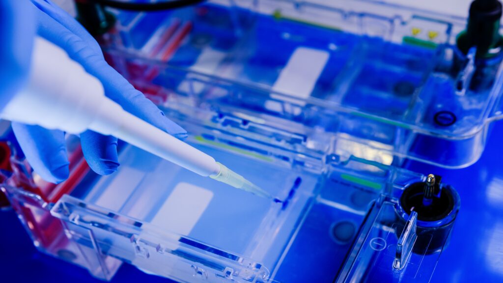
Work on electrophoresis should begin with the preparation of the gel. You can do this yourself or buy a ready-made gel. This is particularly useful for large numbers of separations. Select the type and density of gel to suit your needs and pour it into a mould. In the case of polyacrylamide gels, these are usually special cassettes in which the gel is poured between two plates. In the case of agarose gels, these are flat cuvettes. You can pour a thicker layer and cut the gel into narrower slices later.
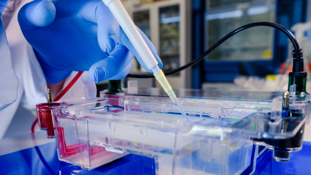
Place the gel in the apparatus and apply the samples to special wells (holes in the gel). After connecting the power supply, the molecules begin to migrate. A dye is often added to the samples to indicate the front of migration. When the dye reaches the end of the gel, the power supply can be turned off and the separation completed.
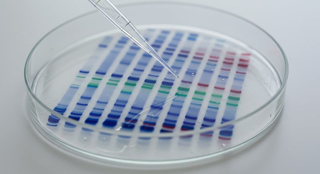
Gel staining
After electrophoresis, the bands (DNA or proteins) are usually invisible. They must be visualised. For this purpose, various dyes are used to make the substances separated on the gel visible.
Protein staining
- Coomassie Brilliant Blue: The most popular, binds with alkaline and aromatic residues. Medium sensitivity.
- Silver staining: Highly sensitive (detects nanograms of protein), but the process is time-consuming and more difficult.
- Fluorescent dyes (eg. SYPRO Ruby): High sensitivity and wide linear range, ideal for quantitative analysis.
DNA/RNA staining
- Ethidium bromide: Incorporates into DNA and fluoresces under UV light.
- SYBR Green/Gold dyes: A safer and often more sensitive alternative to ethidium bromide.
Western Blot
Electrophoresis can also be the first stage of another analysis – Western Blot. In this case, proteins are transferred from the gel to a special membrane. Proteins are detected on the membrane using antibodies.
Analysis of results: Recording and densitometry
The gel image itself is just the beginning. Documentation and analysis systems are used to obtain quantitative data.
- Registration: Gel Doc systems use CCD cameras and appropriate lighting (UV transilluminators or blue/white light) to take a digital photograph of the gel.
- Densitometry: This is the process of measuring the optical density (OD) of bands. The software analyses the intensity of the band’s blackening (or fluorescence). Densitometry is a semi-quantitative technique and allows for a relative assessment of the amount of substances tested in the sample.
Electropherogram analysis also allows the molecular weight or length of nucleic acid to be determined. For this purpose, a reference sample, known as a mass ladder, should be run on the gel. It contains molecules (proteins or nucleic acids) with different molecular weights. The molecular weight can be determined based on the Rf coefficient calibrated against the ladder.
Types of electrophoretic analyses
SDS-PAGE (Sodium Dodecyl Sulphate Polyacrylamide Gel Electrophoresis)
SDS-PAGE is the most common method of protein analysis. The key reagent here is SDS (sodium dodecyl sulphate) – an anionic detergent.
Mechanism of action:
- Denaturation: SDS binds to proteins, causing them to unfold (loss of secondary and tertiary structure). β-mercaptoethanol is often used in addition to reduce disulphide bridges.
- Charging: SDS gives all proteins a strong negative charge. This charge is proportional to the mass of the protein, which “masks” the natural charge of amino acids.
- Separation: This allows proteins to separate in the gel solely on the basis of their molecular weight (size), migrating towards the anode (+).
Isoelectric focusing (IEF)
Isoelectric focusing is a technique for separating proteins based on differences in their isoelectric point (pI).
- Principle of operation: The gel has a fixed pH gradient. The protein migrates in the electric field until it reaches the pH zone equal to its pI.
- Effect: At the pI point, the net charge of the protein is 0, so it stops migrating in the electric field (“focuses”).
- This technique allows the differentiation of protein isoforms that differ by single amino acid residues.
Capillary electrophoresis (CE)
This is a modern, automated version of electrophoresis, in which the process takes place in thin silica capillaries (with a diameter of 25–100 μm).
Advantages:
- Requires very small sample volumes.
- Very high voltage (up to 30 kV) allows for rapid separation.
- High efficiency of Joule heat dissipation.
- Can be coupled with mass spectrometry.
Electroosmotic flow: Electroosmotic flow (EOF) plays an important role here, pulling all ions towards the cathode, regardless of their own charge.
Two-dimensional electrophoresis (2D-PAGE)
2D electrophoresis is a powerful tool in proteomics, combining the two methods described above to separate thousands of proteins from a single sample.
- First dimension (IEF): Separation of proteins according to their isoelectric point (charge).
- Second dimension (SDS-PAGE): The IEF strip is placed on a polyacrylamide gel and separated according to mass.
The result is a ‘map’ of proteins, where each dot represents a unique protein with a specific pI and mass. It is also possible to combine other techniques, e.g. native electrophoresis (first dimension) with reducing electrophoresis (second dimension). In this case, different proteins are separated in the first dimension, and their subunits in the second.
Application
Electrophoresis is ubiquitous in natural sciences and medicine:
- Genetics and Molecular Biology: Testing PCR reaction products, DNA sequencing, genotyping.
- Medical diagnostics: Detection of abnormal haemoglobins (haemoglobinopathies), analysis of blood serum proteins (serum protein electrophoresis) in the diagnosis of myeloma or inflammatory conditions.
- Forensics: DNA profiling (DNA fingerprinting) for the identification of individuals.
- Pharmaceutical industry: Purity control of biological medicines (monoclonal antibodies, vaccines).
Literature
Berg, J. M., Tymoczko, J. L., Gatto, G. J., & Stryer, L. (2018). Biochemia. Wydawnictwo Naukowe PWN.
Hames, B. D. (1998). Gel electrophoresis of proteins: A practical approach. Oxford University Press.
Graphics
Obraz autorstwa wirestock na Freepik
Image by JetalProduções from Pixabay